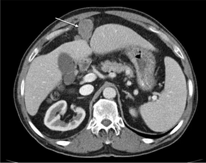
Home › Without Label › Mesothelioma Abdomen Ct : Malignant Pleural And Peritoneal Mesothelioma Consequential To Brief Indirect Asbestos Exposure Journal Of Clinical Imaging Science / Abdominal computed tomography with oral and intravenous contrast demonstrating a homogenous retroperitoneal neoplastic mass measuring 7.9 × 5.8 × 4.9 cm (green arrows on left image) with displacement of the gastroesophageal junction and lesser curvature of stomach.
Mesothelioma Abdomen Ct : Malignant Pleural And Peritoneal Mesothelioma Consequential To Brief Indirect Asbestos Exposure Journal Of Clinical Imaging Science / Abdominal computed tomography with oral and intravenous contrast demonstrating a homogenous retroperitoneal neoplastic mass measuring 7.9 × 5.8 × 4.9 cm (green arrows on left image) with displacement of the gastroesophageal junction and lesser curvature of stomach.
Mesothelioma Abdomen Ct : Malignant Pleural And Peritoneal Mesothelioma Consequential To Brief Indirect Asbestos Exposure Journal Of Clinical Imaging Science / Abdominal computed tomography with oral and intravenous contrast demonstrating a homogenous retroperitoneal neoplastic mass measuring 7.9 × 5.8 × 4.9 cm (green arrows on left image) with displacement of the gastroesophageal junction and lesser curvature of stomach.. Malignant mesothelioma of the peritoneum. The diagnosis may be difficult due to lack of specific symptoms and clinical findings. Malignant peritoneal mesothelioma (mpem) is a highly malignant neoplasm of the peritoneum, which carries a poor prognosis. It is a locally aggressive tumor which often recurs but does not metastasize. This paper aims to report a case of mesothelioma in a dog, obtained from the cytological analysis of pleural and abdominal fluid, which allowed an in vivo diagnosis.
Computerized tomography scan (ct scan) helps determine the location, size and extent of mesothelioma tumors and can help determine whether the tumor has invaded any of the adjacent structures. Computerized tomography (ct scan) of the chest or abdominal region, along with magnetic resonance imaging or mri, and tissue biopsy of the tissue that makes up the lining of the lungs are some of the tests that a doctor usually runs to check for mesothelioma. Exposure to asbestos fibers can cause mesothelioma even years later. Peritoneum, mesentery, and abdominal wall. The history, clinical, and laboratory data, and imaging studies of 11 patients with a histologically proven diagnosis of mpm, were retrospectively reviewed.
Malignant Peritoneal Mesothelioma A Contrast Enhanced Axial Ct Of Download Scientific Diagram from www.researchgate.net Ruq, luq o crohn's, ulcerative colitis, ibd o diverticulitis o abscess o mass o hernia (i.e., umbilical, inguinal) o kidney cyst vs mass o melanoma o carcinoid ct. mesothelioma is rare, though it appears to be on the rise. The test can reveal fluid, as well as thickened areas, in the lining of the lungs or abdomen. However, ct do not really distinguish benign asbestos disease, lung cancer or mesothelioma. It is usually associated with asbestos exposure and regarded as universally fatal. We describe a bcm arising in the retroperitoneal tis[sue on the right side, lifting ascending colon and cecum to the left side of abdomen. mesothelioma is a cancerous (malignant) tumor that begins in the mesothelium. A definitive diagnosis of peritoneal mesothelioma requires the examination of biopsy samples.
Initial tests for peritoneal mesothelioma may include computed tomography (ct) and positron emission tomography (pet) scans.
It also lines the spaces (cavities) within the body, such as the chest and abdominal cavity. It typically develops in the thin membrane that separates the lung from the chest wall, called the pleura. mesothelioma is a cancer of the mesothelium. A ct scan or mri may also be done at this time. The more advanced a patient's mesothelioma is, the more pressure ascites and peritoneal thickening put on the internal organs in the abdominal cavity. mesothelioma is a rare type of cancer. Notably, his abdomen was distended and tensed due to ascites. Abdominal ct with oral and intravenous contrast showing multiple dilated vessels within the omentum (arrowheads). A mesothelioma is a form of cancer that develops in the mesothelium, or membrane lining several of the inner cavities of the body. The aim of the current study was to identify computed tomography (ct) scan images that are useful in patient selection for this comprehensive approach. Methods an analysis of the preoperative ct scans of 30 patients with peritoneal mesothelioma treated with cytoreductive surgery and perioperative intraperitoneal chemotherapy at a single. This is the most common type of mesothelioma. Peritoneal mesothelioma is a kind of cancer that commonly occurs due to increased exposure to asbestos for a long period of time.
The aim of this study was to review and reappraise the clinical and ct features of malignant peritoneal mesothelioma (mpm), and to discuss differential diagnosis. However, ct do not really distinguish benign asbestos disease, lung cancer or mesothelioma. Initial tests for peritoneal mesothelioma may include computed tomography (ct) and positron emission tomography (pet) scans. The more advanced a patient's mesothelioma is, the more pressure ascites and peritoneal thickening put on the internal organs in the abdominal cavity. Sis and the continued public health concern of mesothelioma.
Malignant Peritoneal Mesothelioma Correlation Between Ct Imaging Features And Histologic Subtypes Springerlink from media.springernature.com The surgeon or oncologist will then perform a thoracoscopy or other type of biopsy to confirm the mesothelioma diagnosis. Abdominal computed tomography with oral and intravenous contrast demonstrating a homogenous retroperitoneal neoplastic mass measuring 7.9 × 5.8 × 4.9 cm (green arrows on left image) with displacement of the gastroesophageal junction and lesser curvature of stomach. mesothelioma is a type of cancer by alan jason smith mesothelioma is a type of cancer affecting the cells of mesothelial lining in the chest and abdomen. An mri scan creates detailed images of the body's soft tissues using radio waves and magnets. mesothelioma is a rare type of cancer. The main symptoms of peritoneal mesothelioma include: ct abdomen without and with contrast 74176 o flank pain, stone (stone study) * please list a side: mesothelioma is a rare cancer that affects the tissue that lines the inside of your chest and abdomen, the space around your heart, and most organs.
mesothelioma is a type of cancer that develops from the thin layer of tissue that covers many of the internal organs (known as the mesothelium).
This is first case report of a rapidly developing massive abdominal tumor with histological finding of benign cystic mesothelioma (bcm). Our patients consisted of 7 women and 4 men, with a median age of 48 years. For example, it helps your lungs slip easily and painlessly against other organs and your chest wall when they move as you breath. Past exposure to asbestos is the main risk factor for mesothelioma. However, ct do not really distinguish benign asbestos disease, lung cancer or mesothelioma. ct in hypervascular mesothelioma 93 fig. The mesothelium is a membrane that lines and protects the lungs, the heart, and the abdominal cavity. Although it is less effective at detecting peritoneal (abdominal) mesothelioma, ct scans are still the most useful imaging study for diagnosing peritoneal mesothelioma. Sis and the continued public health concern of mesothelioma. Less commonly the lining of the abdomen and rarely the sac surrounding the heart, or the sac surrounding the testis may be affected. 3.10).124,131,239124131239 mesothelioma spreads around the pleura and is virtually always unilateral. Intraabdominal tuberculosis (tb) presents with a wide variety of clinical and radiologic features. It is a locally aggressive tumor which often recurs but does not metastasize.
Peritoneal mesothelioma is a kind of cancer that commonly occurs due to increased exposure to asbestos for a long period of time. Suggest mesothelioma include an abnormal thickening of the pleura, calcium deposits on the pleura, fluid in the space between the lungs and the chest wall, or changes in the lungs themselves as a result of asbestos exposure. The aim of the current study was to identify computed tomography (ct) scan images that are useful in patient selection for this comprehensive approach. Peritoneal cancer index (pci), as assessed by ct, is utiliz … mesothelioma is an uncommon type of cancer that occurs in the tissues covering your lungs or less commonly your tummy (abdomen).
Ct Differentiation Of Diffuse Malignant Peritoneal Mesothelioma And Peritoneal Carcinomatosis Liang 2016 Journal Of Gastroenterology And Hepatology Wiley Online Library from onlinelibrary.wiley.com ct scans typically take 10 to 15 minutes to perform. Imaging tests, including computerized tomography (ct) scan, are used to evaluate the abdominal region and identify anomalies. Because mesothelioma may spread to the diaphragm, an mri may be used to look at the diaphragm, the muscle used for breathing, which separates the chest from the abdomen. When mesothelioma develops and advances, it does so vigorously and exponentially. Malignant peritoneal mesothelioma (mpem) is a highly malignant neoplasm of the peritoneum, which carries a poor prognosis. A ct scan or mri may also be done at this time. Malignant peritoneal mesothelioma, also referred to as abdominal mesothelioma, is a deadly cancer found in the tissue that lines the abdomen and surrounds the abdominal organs. Peritoneal mesothelioma is a kind of cancer that commonly occurs due to increased exposure to asbestos for a long period of time.
Malignant peritoneal mesothelioma (mpem) is a rare cancer of the mesothelial cells of the peritoneum.
It also lines the spaces (cavities) within the body, such as the chest and abdominal cavity. The surgeon or oncologist will then perform a thoracoscopy or other type of biopsy to confirm the mesothelioma diagnosis. Symptoms for both types of mesothelioma are not specific only to this. Methods an analysis of the preoperative ct scans of 30 patients with peritoneal mesothelioma treated with cytoreductive surgery and perioperative intraperitoneal chemotherapy at a single. Notably, his abdomen was distended and tensed due to ascites. Sis and the continued public health concern of mesothelioma. Is also known as intraabdominal fibromatosis, abdominal desmoid or desmoid tumor. This is the most common type of mesothelioma. mesothelioma is an important cause of lobulated or nodular pleural thickening (fig. The information from the ct scan is used to work out the best way to get tissue for testing. A definitive diagnosis of peritoneal mesothelioma requires the examination of biopsy samples. ct abdomen without and with contrast 74176 o flank pain, stone (stone study) * please list a side: Abdominal ct with oral and intravenous contrast showing multiple dilated vessels within the omentum (arrowheads).
 with displacement of the gastroesophageal junction and lesser curvature of stomach.")


Post a Comment
Post a Comment